Basic principles of genetics

What is a genetic disease?

EXPERTS: Did you know?

The GLA gene provides instructions for making an enzyme alpha-galactosidase A, which is necessary for the metabolism of a lipid called globotriaosylceramide or GL-3. GLA gene mutations prevent alpha-galactosidase A from effectively breaking down GL-3, leading to a buildup of GL-3 in most cells throughout the body. Progressive accumulation of GL-3 causes cell damage, leading to the wide range of symptoms of Fabry disease.20 The condition is a systemic disease that manifests as brain vascular disease, heart disease, and progressive kidney failure.21,23

What is a genetic disease?

­
Genetic diseases result, in whole or in part, by a change in the DNA sequence. Such diseases result when an error or a change in one or more genes leads to a mutant, missing, inactive, or partially active protein, all of which can cause disease. Often when we think about a genetic disease, we’re talking about an
inherited disease.4   


Autosomal-dominant diseases

What is a genetic disease?

­Genetic diseases result, in whole or in part, by a change in the DNA sequence. Such diseases result when an error or a change in one or more genes leads to a mutant, missing, inactive, or partially active protein, all of which can cause disease. Often when we think about a genetic disease, we’re talking about an inherited disease.4  


Autosomal-dominant diseases

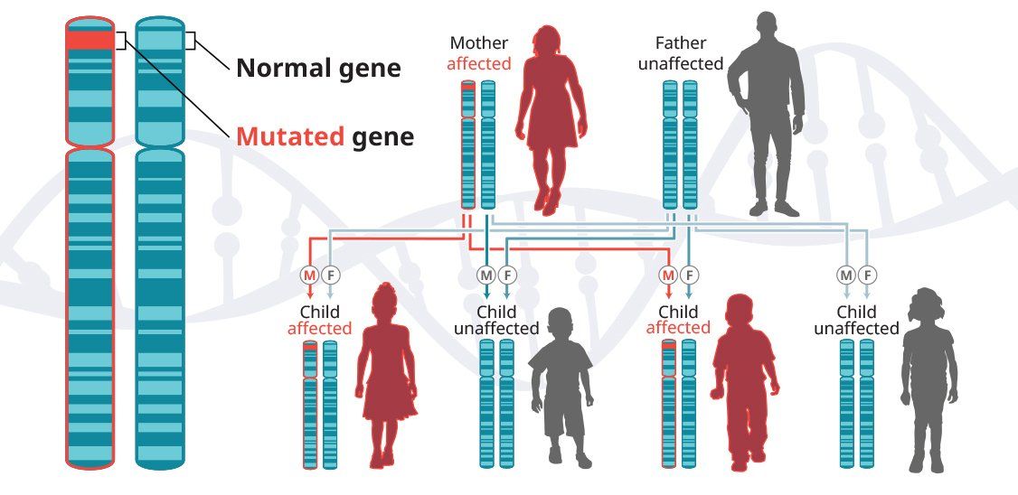
In autosomal-dominant diseases, one copy of the defective gene is sufficient for a person to inherit the condition. In some cases, an affected child inherits the disease from an affected parent.5


Examples of autosomal-dominant diseases


Huntington’s disease (HD)


Huntington’s disease is a progressive neurodegenerative disease that is inherited in an autosomal-dominant pattern, which means that a person inherits one defective copy of the gene and can develop the disease.6-8 When a man or a woman with HD has children, each child has a 50% chance of inheriting the mutated gene and developing the disease. In the United States, approximately 30,000 people have HD and another 200,000 are at risk of developing the disease.9

  • What causes Huntington’s disease?

    Buildup of mutant protein or mutant huntingtin protein (mHTT) is thought to cause HD.#sup_10# The normal function of huntingtin protein is not known, but the mHTT buildup is somehow toxic to brain cells.


    The HTT gene mutation involves a DNA segment known as CAG trinucleotide repeat. CAG is a segment made up of the 3 DNA base pairs, cytosine (C), adenine (A), and guanine (G). In people with HD, the CAG segment repeats 36 to more than 120 times. People with 40 or more CAG repeats will develop HD.#sup_7#


    Repeat expansion mutation


    #img1#


    #ssep#HD first affects the striatum, which is a core structure of the brain. This structure is critical for motor function and reward- and goal-orientated behavior.#sup_6# Loss of brain cells in the striatum leads to the following problems#sup_11-13#: 

    • Motor function issues 

    • Cognitive dysfunction 

    • Psychiatric disturbances#esep#


    Symptoms begin gradually and progress over many years until the death of the individual. The median survival from symptom onset to death is 30 to 40 years.#sup_14#


Spinocerebellar ataxias (SCAs)


Spinocerebellar ataxias (SCAs) are a group of inherited progressive neurodegenerative diseases that mainly affect the cerebellum. Currently, more than 40 types of SCAs have been identified. Not all SCAs are restricted to pure cerebellar degeneration; some types involve other areas of the central nervous system, including the pontine nuclei, spinal cord, cortex, peripheral nerves, and basal ganglia.15


The prevalence of SCAs, which are inherited in an autosomal-dominant fashion, is estimated to be about 1 to 5 in 100,000 individuals.
16  Spinocerebellar ataxia 3 (SCA3) is the most common subtype of type 1 autosomal-dominant ataxias worldwide. The prevalence of SCA3 is estimated to be 1 to 2 in 100,000 people but varies significantly based on geography and ethnicity.17

  • What causes spinocerebellar ataxia 3?

    SCA3, also known as Machado-Joseph disease, is a progressive cerebellar disease that is inherited in an autosomal-dominant pattern.#sup_18,19# Mutations in the ATXN3 gene are thought to cause SCA3.#sup_18#


    The ATXN3 gene provides instructions for how to make ataxin-3, a protein involved in the ubiquitin-proteasome system. This system is responsible for destroying and removing excess or damaged proteins. The molecule ubiquitin attaches to damaged proteins, tagging them for degradation. Ataxin-3 removes ubiquitin from the proteins before they are broken down, so that ubiquitin may be reused. Nonfunctional ataxin-3 cannot remove ubiquitin for reuse, and as a result, ubiquitin, damaged proteins, and ataxin-3 build up and form aggregates within the nucleus of cells. It is not yet well understood how these aggregates affect cell function.#sup_18#


    The ATXN3 gene mutation also involves CAG trinucleotide repeats, similar to other neurodegenerative diseases such as HD. In people with SCA3, the CAG segment repeats more than 50 to 80 times. Usually, a greater number of repeats is associated with earlier onset and faster progression of signs and symptoms, a phenomenon called anticipation.#sup_18#


    Repeat expansion mutation


    #img1#


    #ssep#Brain cells are most affected by mutations in the ATXN3 gene. SCA3 is associated with cell death in the brainstem and cerebellum, as well as the spinal cord. Progressive cell loss in the brain and spinal cord causes the signs and symptoms characteristic of SCA3.#sup_18 #Presenting features include the following#sup_19#:

    • Balance dysfunction

    • Eye muscle movement disorders

    • Gait problems

    • Clumsiness

    • Speech difficulties#esep# 

    Ten to fifteen years after disease onset, individuals usually need assistive devices to help with activities of daily living.#sup_15# The mean survival after disease onset is approximately 21 years.#sup_20#


X-linked recessive diseases

X-linked recessive diseases are caused by mutations in genes on the X chromosome. Men only have one X chromosome, so only one defective copy of the gene is sufficient to cause disease. Women have two X chromosomes, so a mutation would have to occur in both copies of the gene to cause disease; although, women carriers can sometimes display some disease symptoms. It is less likely that women will have two defective copies of this gene. Men are affected by X-linked recessive diseases much more frequently than women. In fact, one characteristic of X-linked inheritance is that fathers cannot pass X-linked traits to their sons.5


Examples of X-linked recessive diseases

 
Fabry disease

 
Fabry disease is caused by mutations in the GLA gene, which is located on the X chromosome, one of two sex chromosomes. Thus, Fabry is an X-linked disease, inherited in a recessive pattern. 

 
Fabry disease occurs in all populations and affects both males and females. It is estimated that there are about 3800 men with Fabry disease in the United States. It is not known how many women are affected by the disease.
22 


Published data from the Fabry registry indicates that men with the disease are expected to live to an average age of 50 to 57 years. Women with Fabry disease are expected to live to an average age of 64 to 72 years.24 

EXPERTS: Did you know?

The GLA gene provides instructions for making an enzyme alpha-galactosidase A, which is necessary for the metabolism of a lipid called globotriaosylceramide or GL-3. GLA gene mutations prevent alpha-galactosidase A from effectively breaking down GL-3, leading to a buildup of GL-3 in most cells throughout the body. Progressive accumulation of GL-3 causes cell damage, leading to the wide range of symptoms of Fabry disease.20 The condition is a systemic disease that manifests as brain vascular disease, heart disease, and progressive kidney failure.21,23

Hemophilia


Hemophilia is a bleeding disorder that interferes with blood clotting. Hemophilia A and hemophilia B are inherited in an X-linked recessive pattern. This means that the genes associated with hemophilia are located on the X chromosome, one of two sex chromosomes.25


For all severities of hemophilia A, it is estimated that this disease occurs in 17.1 cases per 100,000 men. Hemophilia B is not as common. For all severities of hemophilia B, it is estimated that this disease occurs in 3.8 cases per 100,000 men.26 

  • What causes hemophilia?

    Mutations in the F8 gene cause hemophilia A; whereas mutations in the F9 gene cause hemophilia B. A defect or mutation in either of these genes can result in the inability or reduced ability to express blood clotting factor VIII or blood clotting factor IX, or the reduced functionality of one of these proteins. Hemophilia A or B can range from mild to moderate to severe, depending on the level of factor produced. People with mild hemophilia have factor VIII or factor IX levels between 5% to 40% of normal; those with moderate hemophilia have factor levels between 1% to 5% of normal, and those with severe hemophilia have factor levels less than 1% of normal.#sup_27,28#


    When the genes associated with the production of proteins required for blood clotting are missing or mutated, depending on the severity, the patient may bleed spontaneously or may not clot enough to stop bleeding, even after a minor trauma or surgery.#sup_25#


    Current treatment for hemophilia B consists of life-long prophylactic or on-demand injections of replacement factor IX.#sup_29# Despite treatment, people with moderate and severe hemophilia are at risk of bleeds, notably joint bleeds,#sup_30# which can cause chronic inflammation of the synovial membrane and disease of the joints. Intracranial bleeding affects around 2% of people with hemophilia and is potentially life-threatening.#sup_31,32# Appropriate prophylaxis may reduce the risk of bleeding events, including severe intracranial bleeding.#sup_33,34# Although the life expectancy for people with hemophilia has improved, premature mortality remains a challenge in severe hemophilia and/or where access to prophylaxis is limited.#sup_31-33# The average lifespan of people with hemophilia B (United Kingdom, non-HIV) depends on the severity of the disease: 63 years for severe hemophilia and 75 years for mild-to-moderate hemophilia. For comparison, the average lifespan of the general population is 78 years.#sup_32#

Back to top